

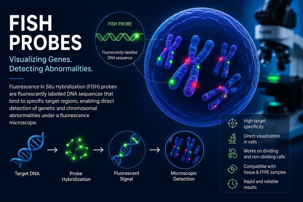
Fluorescence In Situ Hybridization (FISH) is a molecular cytogenetic technique used to detect and visualize specific DNA sequences within cells and tissues. FISH probes are fluorescently labelled DNA sequences designed to bind to complementary target regions, allowing researchers to identify chromosomal abnormalities directly under a fluorescence microscope.
What Are FISH Probes?
FISH probes are short DNA sequences labelled with fluorescent dyes. When a probe binds to its specific target DNA sequence, it produces a fluorescent signal that can be detected and analysed.
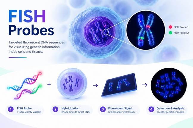
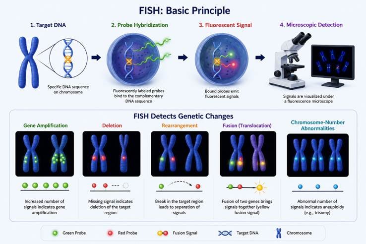
The basic principle is:
Target DNA → Probe Hybridization → Fluorescent Signal → Microscopic Detection
FISH can detect specific genetic changes such as gene amplification, deletion, rearrangement, fusion, and chromosome-number abnormalities.
Types of FISH Probes
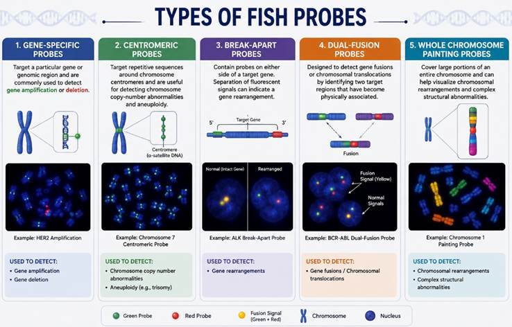
Different probes are designed for different applications:
1. Gene-Specific Probes
Target a particular gene or genomic region and are commonly used to detect gene amplification or deletion.
2. Centromeric Probes
Target repetitive sequences around chromosome centromeres and are useful for detecting chromosome copy-number abnormalities and aneuploidy.
3. Break-Apart Probes
Contain probes on either side of a target gene. Separation of fluorescent signals can indicate a gene rearrangement.
4. Dual-Fusion Probes
Designed to detect gene fusions or chromosomal translocations by identifying two target regions that have become physically associated.
5. Whole Chromosome Painting Probes
Cover large portions of an entire chromosome and can help visualize chromosomal rearrangements and complex structural abnormalities.
Applications of FISH
FISH probes are widely used in:
- Cancer research – gene amplification, deletion, fusion, and rearrangement
- Haematological malignancies – detection of leukaemia- and lymphoma-associated abnormalities
- Genetic disease research – microdeletions, duplications, and aneuploidies
- Prenatal cytogenetics – targeted detection of selected chromosomal abnormalities
- Chromosome research – studying chromosome organization and structural changes
Advantages of FISH
FISH offers several benefits:
- High target specificity
- Direct visualization of genetic abnormalities
- Can be performed on non-dividing cells
- Useful with tissue and FFPE samples
- Relatively rapid targeted analysis
- Provides cellular and chromosomal localization information
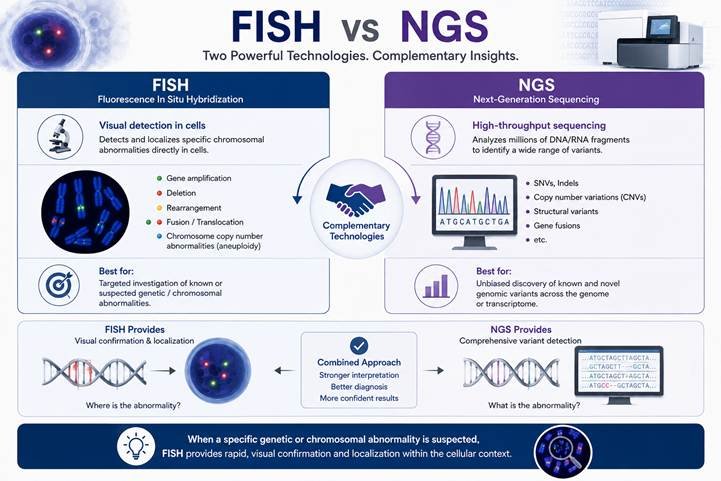
FISH vs NGS
FISH and Next-Generation Sequencing (NGS) provide complementary information. NGS can analyse large numbers of genomic variants, while FISH provides visual confirmation and localization of specific genomic abnormalities within cells.
Therefore, FISH can be particularly valuable when a specific genetic or chromosomal abnormality is suspected.
Conclusion
FISH probes remain an important tool in cytogenetics, cancer research, genetic disease studies, and molecular diagnostics. By enabling targeted visualization of DNA sequences and chromosomal abnormalities, FISH provides valuable information that complements sequencing and other genomic technologies.
Looking for FISH probe-based research or cytogenetic solutions? Choose the probe type according to your target gene, chromosome, or suspected genetic abnormality.