

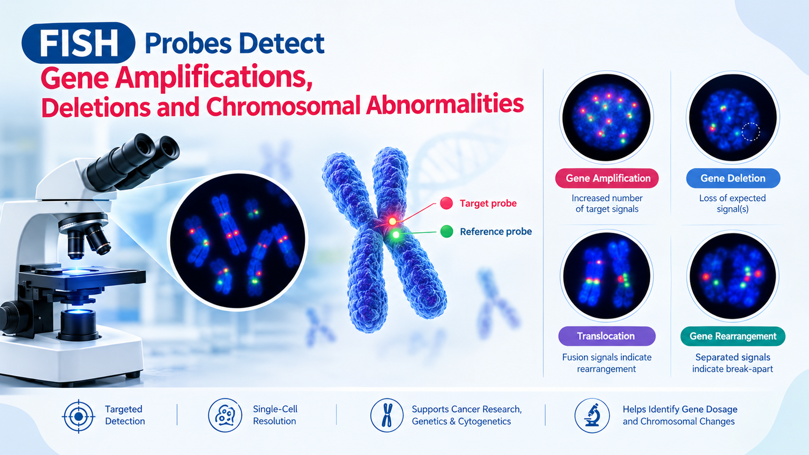
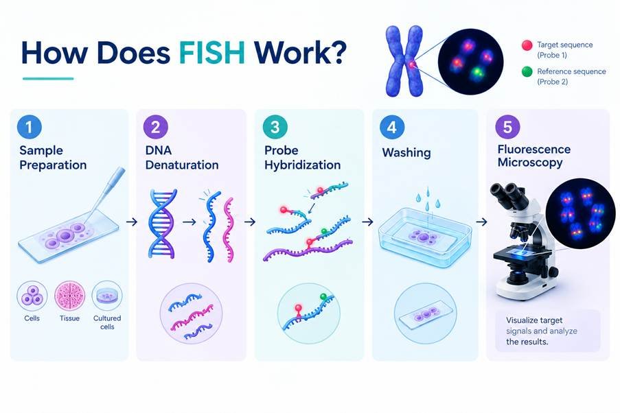
Fluorescence In Situ Hybridization (FISH) is a widely used molecular cytogenetic technique for identifying specific DNA sequences within cells and chromosomes. By using fluorescently labelled FISH probes, researchers can visualize targeted genomic regions under a fluorescence microscope and investigate genetic alterations at the cellular level. FISH is particularly useful for detecting gene amplification, gene deletions, chromosomal abnormalities, gene rearrangements, and copy-number changes. Because it provides targeted information about specific genomic regions, FISH continues to be an important technique in cancer research, genetics, cytogenetics, and molecular biology.
In this article, we explore how FISH probes work and how they help detect gene amplification, deletions, and chromosomal abnormalities.
What Are FISH Probes?
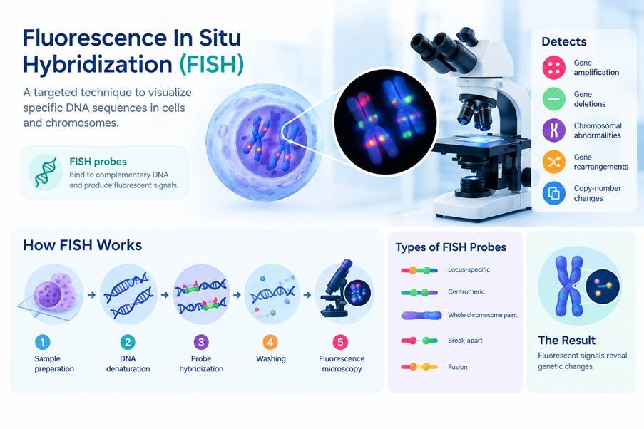
FISH probes are short, labelled DNA sequences designed to bind to a complementary DNA sequence within a chromosome. The probe is tagged with a fluorescent molecule, allowing the target region to be visualized using fluorescence microscopy. Depending on the research objective, different types of probes can be used to investigate specific genomic regions or chromosome-level abnormalities.
Common types of FISH probes include:
- Locus-specific probes: Target a particular gene or genomic region.
- Centromeric probes: Target repetitive DNA sequences around chromosome centromeres and can help assess chromosome copy number.
- Whole chromosome painting probes: Label an entire chromosome and can help visualize chromosomal rearrangements.
- Break-apart probes: Designed to detect rearrangements involving a specific gene.
- Fusion probes: Used to identify specific gene fusion events.
The choice of FISH probe depends on the genomic alteration being investigated.
How Does FISH Work?
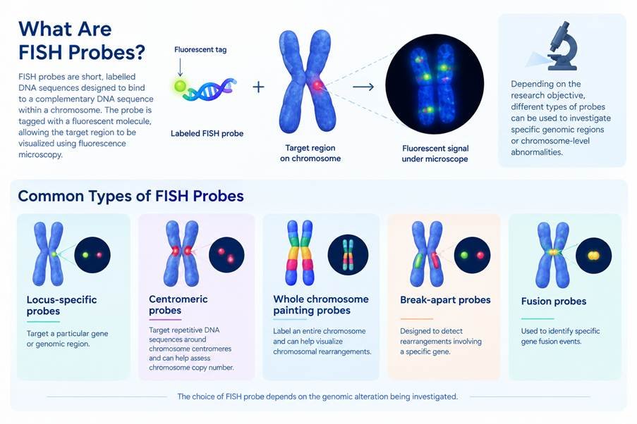
The basic FISH workflow involves hybridizing a fluorescently labelled probe to its complementary DNA sequence in a biological sample.
1. Sample Preparation
Cells or tissue sections are prepared on a microscope slide. Depending on the application, FISH can be performed on various sample types, including cultured cells and tissue specimens.
2. DNA Denaturation
The DNA within the sample is denatured to separate the two DNA strands. This allows the FISH probe to access and bind to its complementary target sequence.
3. Probe Hybridization
The fluorescently labelled FISH probe is introduced to the sample. The probe binds to its complementary DNA sequence through sequence-specific hybridization.
4. Washing
Unbound or non-specifically bound probes are removed through washing steps. This helps improve signal specificity and reduces background fluorescence.
5. Fluorescence Microscopy
The sample is examined under a fluorescence microscope. The fluorescent signals indicate the location and, depending on the probe design and assay, the relative number or arrangement of the targeted sequences.
The observed signal pattern can then be compared with appropriate controls or reference signals.
How FISH Detects Gene Amplification
Gene amplification occurs when multiple copies of a particular genomic region are present within a cell.
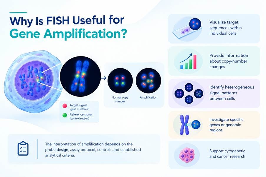
FISH can identify gene amplification by using a probe that specifically targets the gene or genomic region of interest. The number and distribution of fluorescent signals can then be evaluated relative to an appropriate reference. For example, in research involving HER2 amplification, a HER2-specific probe can be evaluated alongside a chromosome 17 reference probe. An increased number of HER2 signals relative to the reference can indicate amplification of the HER2 region. This targeted approach makes FISH particularly useful when researchers need to investigate the copy number of a specific gene.
Why Is FISH Useful for Gene Amplification?
FISH can:
- Visualize target sequences directly within individual cells
- Provide information about copy-number changes
- Identify heterogeneous signal patterns between cells
- Investigate specific genes or genomic regions
- Support cytogenetic and cancer research
The interpretation of amplification depends on the probe design, assay protocol, controls, and established analytical criteria.
How FISH Detects Gene Deletions
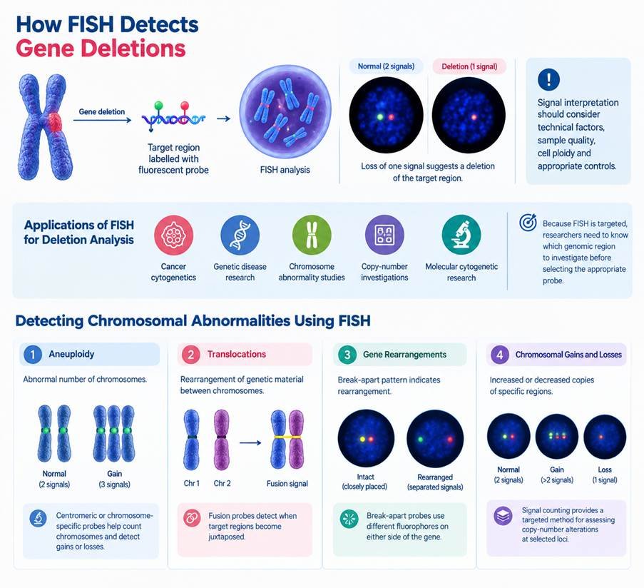
A gene deletion involves the loss of genetic material from a chromosome or genomic region. FISH can be used to investigate deletions by targeting the genomic region of interest with a specific probe. Researchers can compare the observed target signals with reference signals to identify potential loss of the targeted region.
For example, if a locus-specific probe normally produces two signals in a diploid cell but one expected signal is absent, this may indicate a deletion of the corresponding region. However, signal interpretation must consider technical factors, sample quality, cell ploidy, and appropriate controls.
Applications of FISH for Deletion Analysis
FISH-based deletion analysis can be used in:
- Cancer cytogenetics
- Genetic disease research
- Chromosome abnormality studies
- Copy-number investigations
- Molecular cytogenetic research
Because FISH is targeted, researchers generally need to know which genomic region they want to investigate before selecting the appropriate probe.
Detecting Chromosomal Abnormalities Using FISH
Chromosomal abnormalities can involve changes in chromosome number or structure. FISH probes can help investigate several types of chromosomal alterations.
1. Aneuploidy
Aneuploidy refers to an abnormal number of chromosomes. Centromeric or chromosome-specific FISH probes can be used to count signals corresponding to particular chromosomes. This can help researchers identify potential gains or losses of chromosomes.
2. Translocations
A chromosomal translocation occurs when genetic material is rearranged between chromosomes. Fusion probes can be designed to target two genes or genomic regions involved in a known rearrangement. When the target regions become juxtaposed, the resulting fluorescent signal pattern can indicate a potential fusion event.
3. Gene Rearrangements
Break-apart FISH probes can be used to investigate rearrangements involving a particular gene. Typically, probes labelled with different fluorophores bind to regions on either side of the target gene. In an intact region, the signals appear together or in close proximity. A rearrangement can separate these signals, producing a characteristic break-apart pattern.
4. Chromosomal Gains and Losses
FISH can also help investigate increased or decreased copies of specific chromosome regions. Signal counting provides a targeted method for assessing copy-number alterations at selected loci.
Common FISH Probe Designs and Their Applications
| FISH Probe Type | Primary Application |
| Locus-specific probe | Detection of specific genes or genomic regions |
| Centromeric probe | Chromosome enumeration and copy-number assessment |
| Whole chromosome painting probe | Investigation of chromosome-level rearrangements |
| Break-apart probe | Detection of gene rearrangements |
| Fusion probe | Detection of specific gene fusion events |
Selecting the appropriate probe design is essential for obtaining meaningful FISH results.
FISH Signal Patterns: What Do They Indicate?
The interpretation of FISH results depends on the specific probe and experimental design. Common patterns may include:
Normal signal pattern:
The expected number and arrangement of signals are observed for the targeted region.
Increased signal count:
An increased number of target signals may indicate a gain or amplification, depending on the assay design.
Reduced signal count:
A loss of an expected signal may indicate a deletion or loss of the targeted region.
Separated signals:
With break-apart probes, separation of differently labelled signals can indicate a potential rearrangement.
Colocalized or fusion signals:
With fusion probes, overlapping signals can indicate proximity or fusion of targeted regions.
Importantly, FISH results should not be interpreted based on signal patterns alone. Appropriate controls, probe specifications, sample quality, imaging conditions, and validated interpretation criteria are important for reliable analysis.
FISH in Cancer Research
FISH has an important role in cancer cytogenetics because cancer cells can acquire genomic alterations such as gene amplification, deletion, translocation, and chromosome-number changes.
Researchers can use FISH to investigate specific alterations associated with:
- Tumour biology
- Gene amplification
- Oncogenic rearrangements
- Chromosomal instability
- Copy-number alterations
- Molecular classification of cancer
For example, FISH-based analysis of specific genomic alterations can complement other molecular techniques used in cancer research.
FISH vs Conventional Karyotyping
Both FISH and karyotyping are used in cytogenetic research, but they provide different levels of information.
| Feature | FISH | Conventional Karyotyping |
| Detection approach | Targeted | Genome-wide chromosome-level |
| Resolution | Higher for selected regions | Lower for small alterations |
| Target required beforehand | Generally yes | No specific target required |
| Analysis | Fluorescence microscopy | Chromosome visualization |
| Small targeted alterations | Can detect selected changes | May not resolve smaller changes |
| Whole-genome overview | Limited | Provides chromosome-level overview |
FISH is therefore particularly useful when researchers have a specific genomic region or chromosomal abnormality they want to investigate.
Advantages of FISH Probes
FISH offers several advantages for molecular cytogenetic research:
Targeted Detection
FISH probes can focus on specific genes, loci, chromosomes, or rearrangements.
Single-Cell Visualization
Signals can be visualized within individual cells, allowing researchers to examine cellular heterogeneity.
Specificity
Sequence-specific hybridization allows targeted investigation of genomic regions.
Versatile Applications
Different probe designs enable investigation of amplification, deletion, copy-number changes, and rearrangements.
Compatibility with Cytogenetic Research
FISH can complement conventional cytogenetics and other molecular approaches.
Limitations of FISH
Despite its advantages, FISH also has limitations. Because FISH is generally a targeted technique, researchers typically need prior knowledge of the genomic region or abnormality they want to investigate. It is therefore not a replacement for genome-wide sequencing approaches when the objective is to discover unknown variants across the genome.
Other factors that can influence FISH analysis include:
- Probe design
- Sample quality
- Hybridization efficiency
- Background fluorescence
- Signal quality
- Microscopy conditions
- Appropriate controls
- Interpretation criteria
For comprehensive genomic profiling, FISH may be used alongside techniques such as next-generation sequencing (NGS), microarray analysis, or chromosomal microarray depending on the research objective.
Applications of FISH Probes in Molecular Biology
FISH technology has applications across several research areas, including:
- Cancer research
- Cytogenetics
- Genetic disorder research
- Chromosomal abnormality studies
- Gene amplification research
- Gene deletion analysis
- Gene fusion studies
- Copy-number analysis
- Molecular diagnostics research
The specific application determines the type of probe, sample preparation method, imaging approach, and interpretation strategy.
Conclusion
FISH probes provide a targeted and visually informative method for investigating specific genomic regions within cells. By using appropriately designed probes, researchers can study gene amplification, deletions, chromosome-number changes, gene rearrangements, and other chromosomal abnormalities. The ability to visualize specific DNA sequences using fluorescence microscopy makes FISH an important tool in molecular cytogenetics, cancer research, genetics, and biomedical research. For reliable results, selecting the appropriate FISH probe design, maintaining sample quality, and using suitable controls are essential. FISH can also be integrated with other genomic technologies to provide complementary information about genetic and chromosomal alterations.