

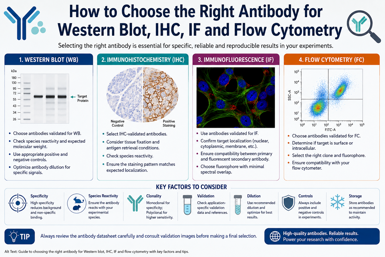
Choosing the right antibody is an important step for obtaining reliable and reproducible results in biomedical research. Antibodies are widely used in techniques such as Western blot (WB), immunohistochemistry (IHC), immunofluorescence (IF), and flow cytometry (FC) to detect and study specific proteins.
However, an antibody that performs well in one application may not necessarily work effectively in another. Therefore, researchers should consider factors such as application validation, target specificity, species reactivity, clonality, host species, fluorophore, and sample preparation before purchasing an antibody.

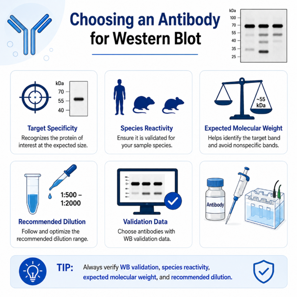
Choosing an Antibody for Western Blot
Western blot is commonly used to identify and compare protein expression in biological samples such as cell or tissue lysates.
When selecting an antibody for Western blot, the most important factor is application-specific validation. Look for antibodies that have been tested and validated for WB.
Important factors include:
Target specificity:
The antibody should specifically recognize the protein of interest and ideally produce a band at the expected molecular weight.
Species reactivity:
Check whether the antibody has been validated for your sample species, such as human, mouse, rat, or other organisms.
Expected molecular weight:
Knowing the expected molecular weight of the target helps in interpreting the Western blot and identifying potential nonspecific bands.
Recommended dilution:
Follow the manufacturer’s recommended dilution range and optimize it according to your experimental conditions.
Validation data:
Antibodies supported by experimental WB images and validation information can provide greater confidence during selection.
Tip: Do not choose an antibody only because it targets the correct protein. Always verify WB validation, species reactivity, expected molecular weight, and recommended dilution.
Choosing an Antibody for Immunohistochemistry (IHC)
Immunohistochemistry is used to detect and visualize proteins in tissue sections. Antibody selection for IHC requires additional consideration because tissue fixation and antigen retrieval can affect epitope accessibility.
Key factors to consider:
IHC validation:
Choose an antibody that has been specifically validated for IHC.
Tissue species:
Confirm that the antibody recognizes the species from which your tissue sample is obtained.
Fixation:
Common fixation methods include formalin fixation and paraformaldehyde fixation. Different fixation conditions can influence antibody binding.
Antigen retrieval:
Some epitopes may become masked during fixation. Antigen retrieval methods, such as heat-induced epitope retrieval or enzymatic treatment, may be required.
Localization:
The expected cellular or tissue localization of the target should be considered when interpreting staining patterns.
A properly validated IHC antibody can help produce clear and biologically meaningful staining while reducing nonspecific background.
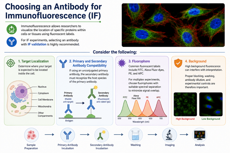
Choosing an Antibody for Immunofluorescence (IF)
Immunofluorescence allows researchers to visualize the location of specific proteins within cells or tissues using fluorescent labels.
For IF experiments, selecting an antibody with IF validation is highly recommended.
Consider the following:
Target localization:
Determine whether your target is expected to be located in the nucleus, cytoplasm, cell membrane, mitochondria, or another cellular compartment.
Primary and secondary antibody compatibility:
If using an unconjugated primary antibody, the secondary antibody must recognize the host species of the primary antibody.
For example:
Primary: Rabbit anti-target antibody
Secondary: Fluorescent anti-rabbit IgG
Fluorophore:
Common fluorescent labels include FITC, Alexa Fluor dyes, PE, and APC. For multiplex experiments, choose fluorophores with suitable spectral separation to minimize signal overlap.
Background:
High background fluorescence can interfere with interpretation. Proper blocking, washing, antibody dilution, and experimental controls are therefore important.
Choosing an Antibody for Flow Cytometry
Flow cytometry is used to analyze proteins on individual cells and can measure multiple markers simultaneously.
Antibody selection for flow cytometry requires special attention to clone, fluorophore, target location, and instrument compatibility.
Surface vs intracellular targets
First determine whether your target is present on the cell surface or inside the cell.
Surface markers can generally be detected without permeabilization, while intracellular proteins usually require fixation and permeabilization before antibody staining.
Clone selection
Several antibody clones may be available against the same target. Different clones can have different binding characteristics and performance.
Therefore, researchers should review available validation data and select a clone appropriate for their application.
Fluorophore selection
Common flow cytometry fluorophores include:
- FITC
- PE
- APC
- PerCP
- PE-Cy7
- APC-Cy7
- Brilliant Violet dyes
The selected fluorophore must be compatible with the lasers and detectors of your flow cytometer.
This becomes particularly important when designing multicolor flow cytometry panels.
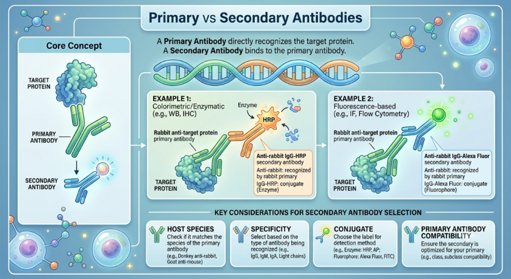
Primary vs Secondary Antibodies
A primary antibody directly recognizes the target protein, while a secondary antibody binds to the primary antibody.
For example:
Primary: Rabbit anti-target protein
Secondary: Anti-rabbit IgG-HRP
For fluorescence-based applications:
Primary: Rabbit anti-target protein
Secondary: Anti-rabbit IgG-Alexa Fluor
When selecting a secondary antibody, check its host species, specificity, conjugate, and compatibility with the primary antibody.
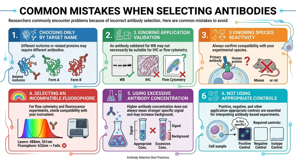
Common Mistakes When Selecting Antibodies
Researchers commonly encounter problems because of incorrect antibody selection. Some common mistakes include:
1. Choosing only by target name
Different isoforms or related proteins may require different antibodies.
2. Ignoring application validation
An antibody validated for WB may not necessarily be suitable for IHC or flow cytometry.
3. Ignoring species reactivity
Always confirm compatibility with your experimental species.
4. Selecting an incompatible fluorophore
For flow cytometry and fluorescence experiments, check compatibility with your instrument.
5. Using excessive antibody concentration
Higher antibody concentration does not always mean stronger specific signal and may increase background.
6. Not using appropriate controls
Positive, negative, and other application-appropriate controls are essential for interpreting antibody-based experiments.
Conclusion
Choosing the right antibody is essential for achieving specific, reliable, and reproducible experimental results. The ideal antibody depends not only on the target protein but also on the application, species, sample preparation, clonality, and validation data.
For Western blot, focus on WB validation, specificity, and expected molecular weight. For IHC, consider tissue fixation, antigen retrieval, and staining validation. For immunofluorescence, pay attention to protein localization, fluorophore selection, and antibody compatibility. For flow cytometry, consider clone, target location, fluorophore, and instrument compatibility.
Before purchasing, always review the manufacturer’s datasheet and application-specific validation data. A carefully selected antibody can significantly improve the quality and reproducibility of your research.